EYE FIELD /27 OCTOBER 2026
Uneven Eyes in Photos: Why the Eye Score Changes
Uneven eyes in a photograph and a changed eye score are not the same finding. Our eye field compares detected geometry in the input image; it cannot identify why someone’s eyes look different. In our existing one-portrait records, the eye reading ranged from 98.0 to 99.1 after file changes, without a new photograph of the person.
Why can the two eyes look different in a photo?
Start with the photograph: the camera view, the light across the image, the distance and the saved file can differ between pictures. Our five-input comparison isolates prepared file changes, not physical causes of uneven eyes. It does not tell us how your eyes look outside that photograph.
Angle: distinguish a rotated file from a turned face
Our +10° flat rotation changed the eye reading from 98.0 to 99.1: +1.1 points, repeated three times. The total changed from 98.0 to 98.1. This was an in-plane rotation with interpolation, not a head turn. It shows that the displayed eye result can change with image preparation; it does not measure the effect of turning your head.
Light: our test changed tone, not the lamp
Gamma 1.4 darkening changed the eye field from 98.0 to 98.1: +0.1 points in each of three uploads. The total fell to 97.6. This synthetic edit did not move a light source or test uneven lighting across the face, so it cannot quantify that capture effect.
Distance: resizing is not stepping away
The 256×256 LANCZOS copy returned 98.5 for eyes, +0.5 points on all three uploads, with a total of 98.1. That condition changed resolution, not camera distance. For actual near and far photographs, our angle-and-distance record keeps the capture conditions separate. These five files alone cannot assign an eye-score difference to distance.
Compression: the eye field and total can disagree
The JPEG quality 20 copy with subsampling 2 returned 98.6 for eyes, +0.6 points, while the total fell from 98.0 to 97.5: −0.5 points. All three uploads matched at the displayed precision. A higher eye reading after compression does not mean that the eyes became more alike or that the compressed copy is more accurate.
When comparing photographs, keep the camera position, distance, expression and lighting as consistent as possible, and retain the original files. When comparing repeated uploads, use the exact same file. These records describe photo-based geometry; they do not establish a physical cause or a treatment.
The eye field is the question here
This page examines the eye component of our 0–100 photo index, rather than turning the whole-face total into a statement about eyes. The same photographed person appears in every file below. We did not collect clinical eye measurements or inspect the underlying landmark coordinates.
The source observations were recorded on 26 October 2026. We reused them for this page on 27 October; these are not new trials. Each file was uploaded three times in one Chrome session. The separate unchanged-file repeat check also shown below used another tab.
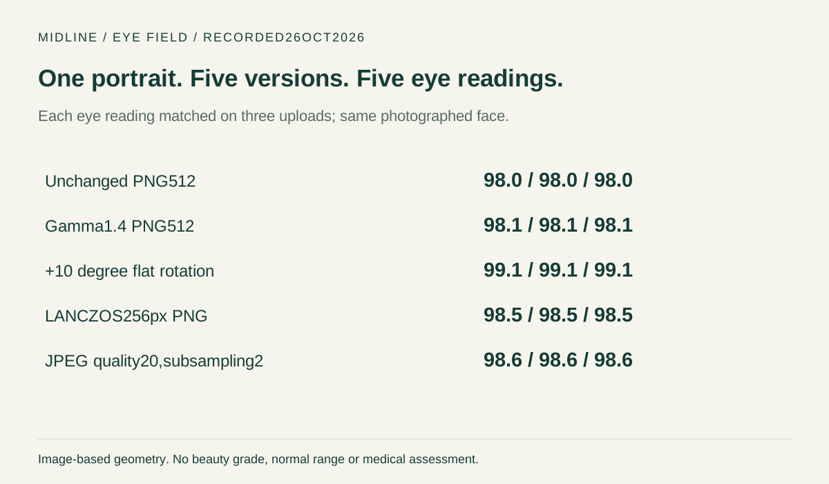
Every original eye reading, with the total beside it
| Date | Input | Repeat | Eyes | Eye minus baseline | Overall |
|---|---|---|---|---|---|
| 2026-10-26 | Unchanged PNG 512 | 1 | 98.0 | +0.0 | 98.0 |
| 2026-10-26 | Unchanged PNG 512 | 2 | 98.0 | +0.0 | 98.0 |
| 2026-10-26 | Unchanged PNG 512 | 3 | 98.0 | +0.0 | 98.0 |
| 2026-10-26 | Gamma 1.4 PNG 512 | 1 | 98.1 | +0.1 | 97.6 |
| 2026-10-26 | Gamma 1.4 PNG 512 | 2 | 98.1 | +0.1 | 97.6 |
| 2026-10-26 | Gamma 1.4 PNG 512 | 3 | 98.1 | +0.1 | 97.6 |
| 2026-10-26 | +10 degree flat rotation | 1 | 99.1 | +1.1 | 98.1 |
| 2026-10-26 | +10 degree flat rotation | 2 | 99.1 | +1.1 | 98.1 |
| 2026-10-26 | +10 degree flat rotation | 3 | 99.1 | +1.1 | 98.1 |
| 2026-10-26 | LANCZOS 256 px PNG | 1 | 98.5 | +0.5 | 98.1 |
| 2026-10-26 | LANCZOS 256 px PNG | 2 | 98.5 | +0.5 | 98.1 |
| 2026-10-26 | LANCZOS 256 px PNG | 3 | 98.5 | +0.5 | 98.1 |
| 2026-10-26 | JPEG quality 20,subsampling 2 | 1 | 98.6 | +0.6 | 97.5 |
| 2026-10-26 | JPEG quality 20,subsampling 2 | 2 | 98.6 | +0.6 | 97.5 |
| 2026-10-26 | JPEG quality 20,subsampling 2 | 3 | 98.6 | +0.6 | 97.5 |
The unchanged 512×512 PNG produced an eye reading of 98.0 on all three uploads. Gamma 1.4 darkening returned 98.1 each time:98.1−98.0=+0.1 points. This was a synthetic tone edit; it does not reproduce physical directional illumination.
The flat+10-degree rotated file returned 99.1 on all three uploads:99.1−98.0=+1.1 points. It used BICUBIC interpolation, a fixed 512×512 canvas and white fill. It was not a three-dimensional head turn, and we did not separate interpolation from the other preparation effects.
The 256×256 LANCZOS-resampled PNG returned 98.5 three times, or 98.5−98.0=+0.5. The 512×512 JPEG, encoded at quality 20 with subsampling 2, returned 98.6 three times, or 98.6−98.0=+0.6. A smaller or more compressed input receiving a higher eye reading does not establish a more accurate measurement.
Why the total does not answer an eye question
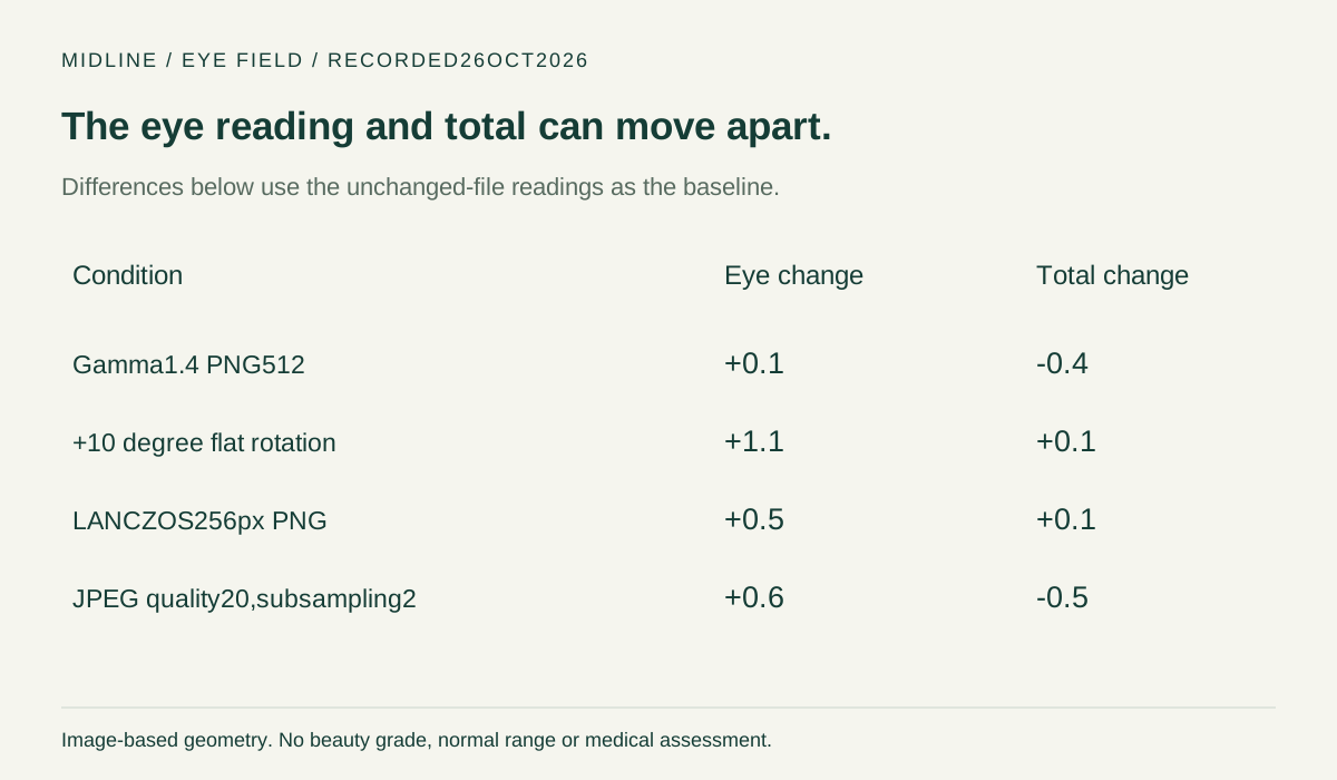
For the JPEG copy, the eye field rose by 98.6−98.0=+0.6 while the overall score fell by 97.5−98.0=−0.5. For the darker file, eyes rose by 0.1 while the total fell by 0.4. Reading only the total would describe those eye-field movements incorrectly.
The rotation changed the eye field by 1.1 points but the total by 0.1. That contrast belongs to this input set. It does not establish that rotation always raises an eye score, or that a higher eye score means a person looks better.
What repeated uploads did—and did not—settle
Our separate 26 October check uploaded the exact unchanged baseline ten times. Every eye reading was 98.0. The displayed eye-field repeat range was max(98.0,…,98.0)−min(98.0,…,98.0)=0.0 across those ten uploads. Each nonzero eye difference above exceeds that observed display range.
An observed 0.0 range at one decimal place is not proof of zero detector noise or anatomical accuracy. It supplies no normal eye-asymmetry cutoff and no statistical significance test. We did not record raw eye-landmark coordinates, so these runs cannot tell which point moved or whether a small coordinate change was hidden by rounding.
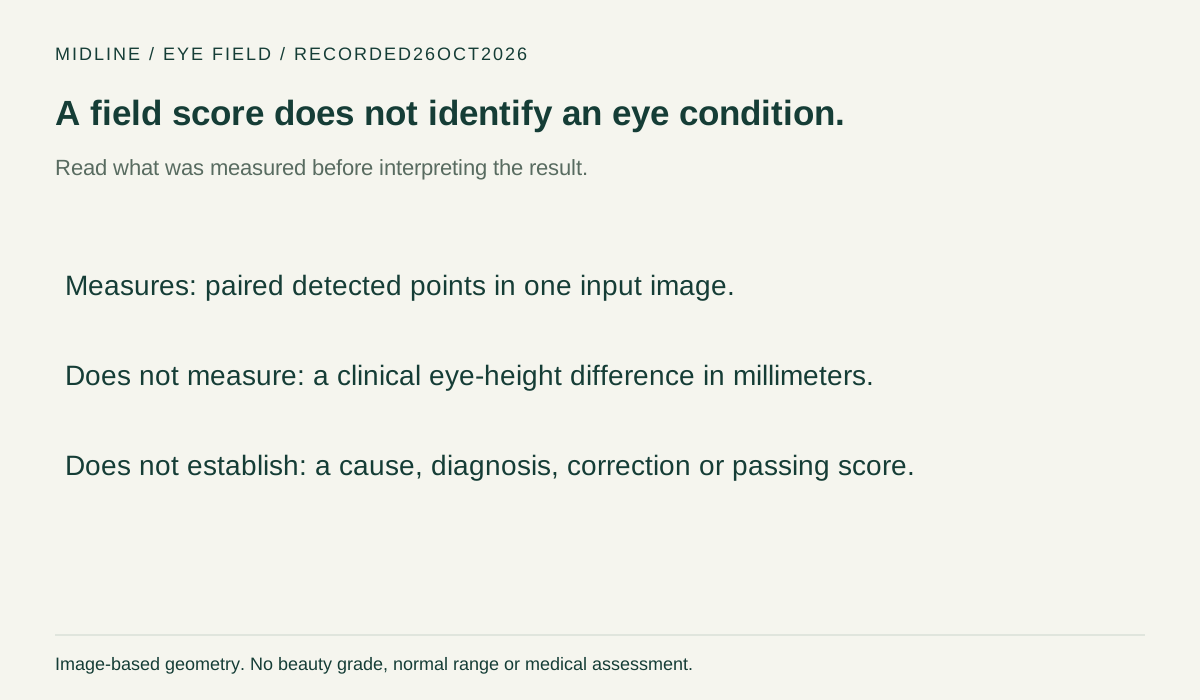
Use the eye result without turning it into a diagnosis
Open the face symmetry test with one clear front-facing photograph. Inspect the overlay and read the eye field alongside the total. For a repeated-file comparison, keep the exact input rather than making a smaller or color-edited copy. A refusal should stay recorded as a refusal, not a low score.
The actionable limit is simple: this output is tied to a file and a detector. It is not a measurement of eyelid health, visual function or a cause of uneven eyes. This page offers no correction routine or treatment recommendation.
Questions about uneven eyes in photos
Does 99.1 mean my eyes are clinically normal?
No clinical normality scale was established by these measurements.99.1 is the output for one prepared file, not a health judgment.
Can I use the total instead of the eye field?
The JPEG case shows why that can mislead: eyes increased 0.6 points while the total decreased 0.5. These were separately recorded outputs.
Did the person’s eyes change between the five files?
The variants were derived from a fixed portrait, not new captures. The observed changes are image-processing outputs; they do not establish a physical change.
Replay these records
All original readings and the ten-repeat check · The full field-by-field comparison. This page reuses our own dated records; no outside measurement numbers were inserted.